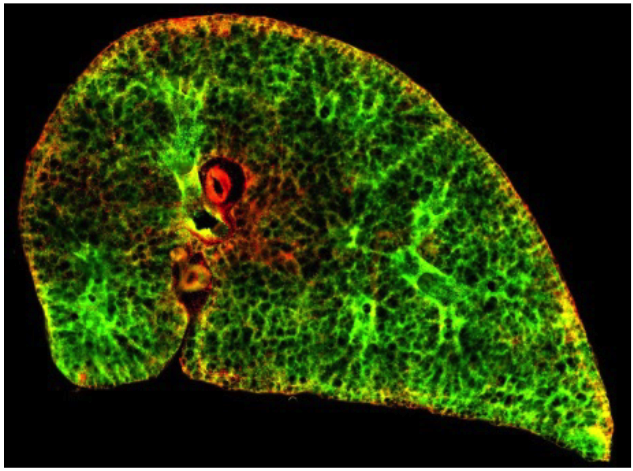
Live imaging of alveologenesis in precision-cut lung slices reveals dynamic epithelial cell behaviour

Researchers in the Dean lab have published exciting exciting novel insights into alveoli development. Alveoli are the site of gas exchange in the lungs. Damage to alveoli, is a component of many chronic and acute lung diseases such as emphysema and Idiopathic pulmonary fibrosis. In addition, insufficient generation of alveoli results in bronchopulmonary dysplasia, which affects many babies born prematurely. Visualising the process of alveolar development (alveologenesis) is important so that we can begin to explore potential ways to repair and regenerate lung tissue. Because the lungs are situated deep inside the body, it is difficult to see alveologenesis happening in the body. In this study, we have established a new method to visualise this process live, in slices of lung tissue. This is the first time that we have been able to see the process of alveologenesis in real-time. Our study finds that during alveologenesis, a key type of cells, epithelial cells are highly mobile and we have identified how epithelial cells associate with each other to form new alveoli. We also show that by adding drugs to the slices, we can interfere with the process of alveologenesis. Our study has established a new system that enables us to conduct research on how alveoli form and to test potential treatments to repair damaged alveoli.
Tweet link :
https://twitter.com/ImperialNHLI/status/1105450997546131456
Paper link:
https://rdcu.be/bqC71
Akram, K. M., Yates, L. L., Mongey, R., Rothery, S., Gaboriau, D. C. A., Sanderson, J., Hind, M., Griffiths, M. & Dean, C. H. 2019 (published 12 March). Nature Communications 10, Article number: 1178
